
Quick Links
Vitreo-retinal Services
The retina is a delicate, light-sensitive layer at the back of the eye responsible for transmitting visual information to the brain. The vitreous is the clear gel that fills the eye’s interior and helps maintain its shape. Together, these structures play a critical role in vision. Vitreo-retinal diseases refer to a wide range of conditions that affect the retina, vitreous, or both, potentially leading to permanent vision loss if left untreated.
These conditions can arise due to aging, diabetes, injury, retinal detachment, or vascular blockages, and may present suddenly or develop gradually. At Chakrabarthi Eye Care Centre, our specialized retina unit is equipped to diagnose and manage even the most complex retinal disorders using cutting-edge diagnostics and advanced surgical interventions.
Read More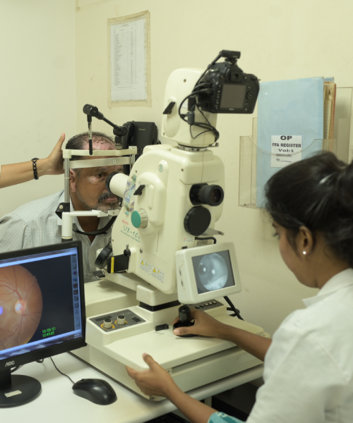
Common Vitreo-Retinal Conditions
Diabetic Retinopathy
A diabetes-related condition that damages the retina's blood vessels, leading to leakage, swelling, or abnormal growth. It may cause floaters, blurred vision, or even blindness. Regular screening is key to early detection.
Diabetic retinopathy with clinically significant macular edema requires
- Strict diabetes control
- Combination of grid laser photocoagulation and intravitreal injection of an anti vegf antibody/sustained release steroid implant
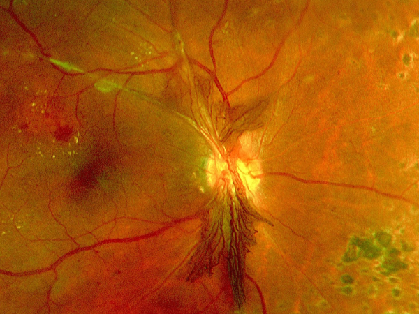
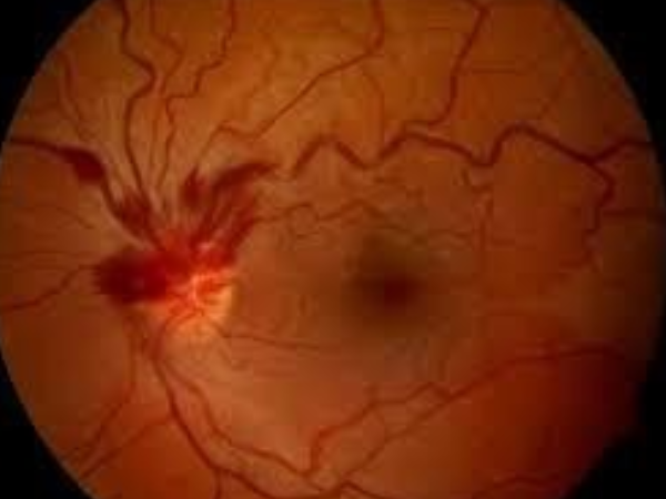
In a poorly controlled diabetic who has not been screened for diabetic retinopathy Plaques of yellow cholesterol accumulation under the macula with severe retinal swelling will cause gross irreversible visual loss You need an eye check up with retinopathy screening tests within one year of detection of diabetes
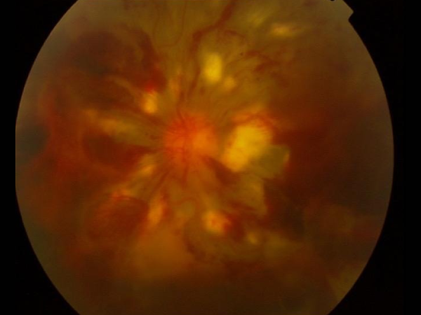
An untreated case of Diabetic Retinopathy with late diagnosis has become An Advanced Diabetic eye disease with Proliferative retinopathy, Preretinal and vitreous haemorrhage and Tractional retinal detachment. This stage requires a major vitreoretinal surgery with silicone oil tamponade and another procedure later on to remove the silicone oil
Retinal Detachment
Occurs when the retina lifts away from the eye’s back surface. Warning signs include sudden floaters, light flashes, or a curtain-like shadow. It’s an emergency that needs immediate surgical attention.
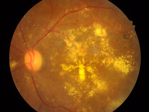
Age-Related Macular Degeneration (AMD)
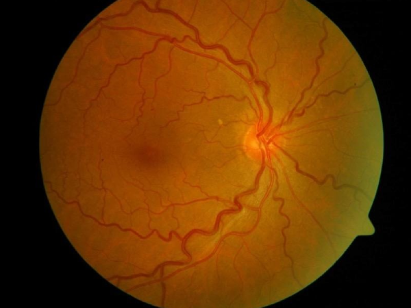
A condition affecting the central retina (macula), leading to gradual loss of central vision. It occurs in dry and wet forms, with the latter requiring urgent injection therapy.
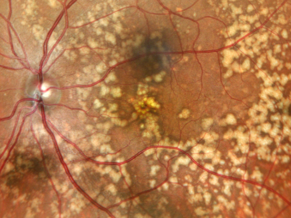
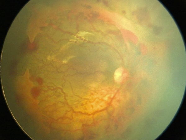
Age related macular degeneration with drusen and pigmentary atrophy
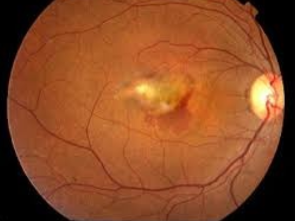
Wet ARMD with subretinal bleed
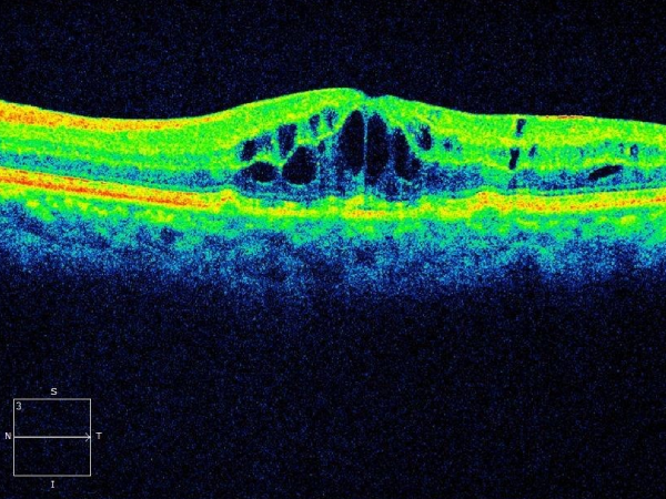
Macular Hole
A small opening in the macula causing blurred or distorted central vision. Surgery, such as vitrectomy, is often needed to restore sight.
Retinal Vein Occlusion (RVO)
A blockage in the retinal veins that can cause swelling, hemorrhage, and sudden vision loss. Treated with injections, laser, or implants.
Incomplete Occlusio
Non-Ischaemic Vein Occlusion
Ischaemic Vein Occlusion
Hemi Retinal Vein Occlusion
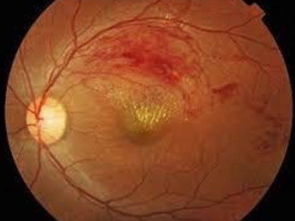
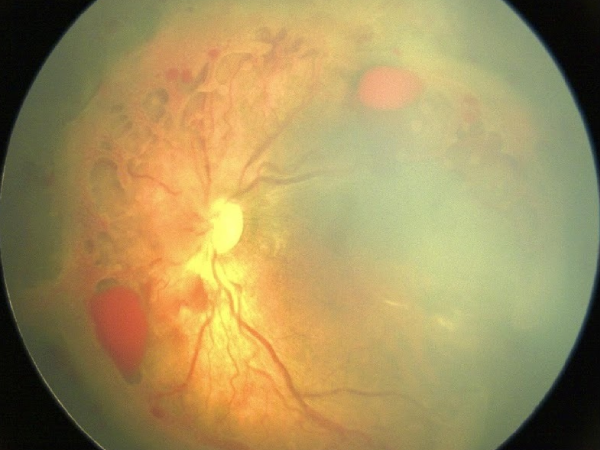
Branch retinal vein occlusion with collaterals and macular edema
BRVO post sectorial laser photocoagulation
ISCHAEMIC
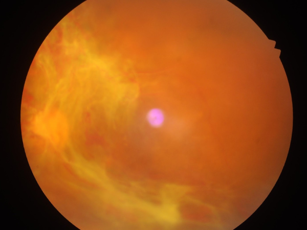
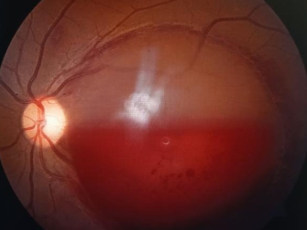
Subhyaloid haemorrhage in a pregnant lady in Fist trimester pregnancy following a bout of vomiting
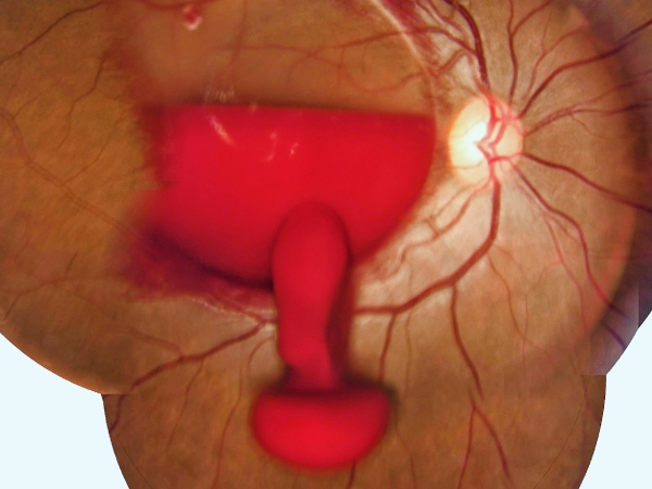
Laser hyaloidotomy to drain subhyaloid blood in a patient with a retinal macroaneurysm
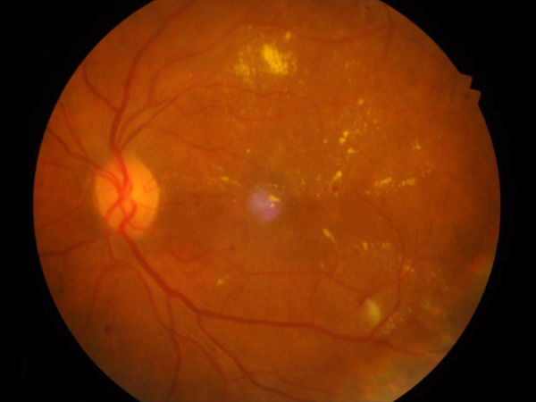
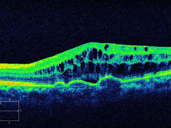
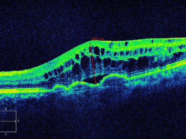
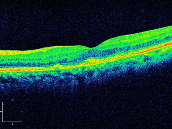
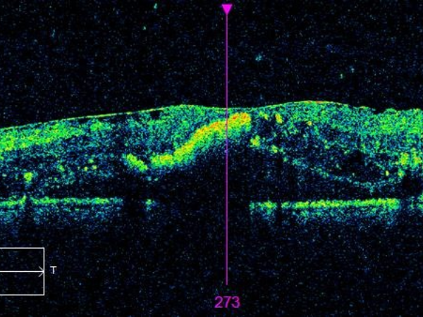
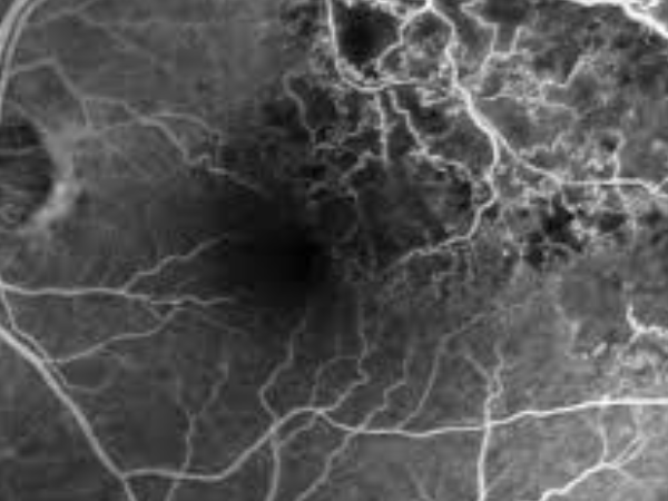
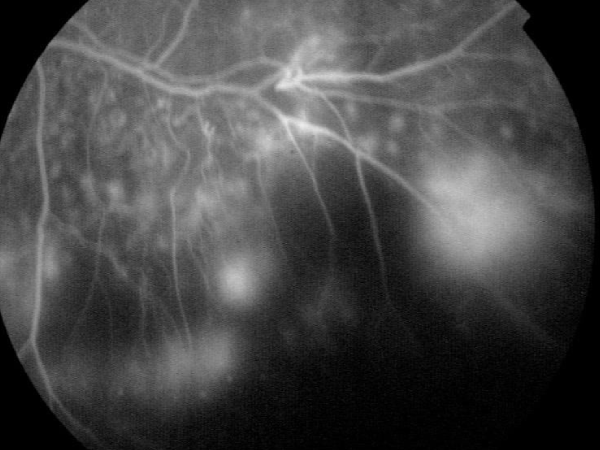
Central Serous Retinopathy
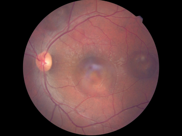
Fundus
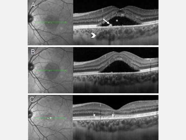
OCT
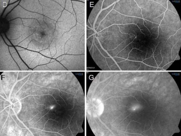
Fluorescein Fundus Angiography
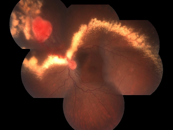
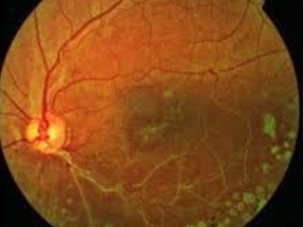
Retinal Angioma with Exudation
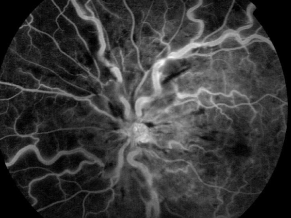
Cilioretinal Artery Occlusion
Central Retinal Artery Occlusion with Cilioretinal Artery Sparing
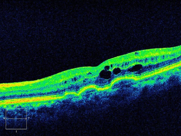
Epiretinal Membrane (Macular Pucker)
A thin membrane on the retina’s surface that distorts vision. While mild cases need monitoring, significant distortion may need surgical removal.
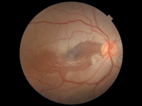
Pigment epithelelitis following viral fever
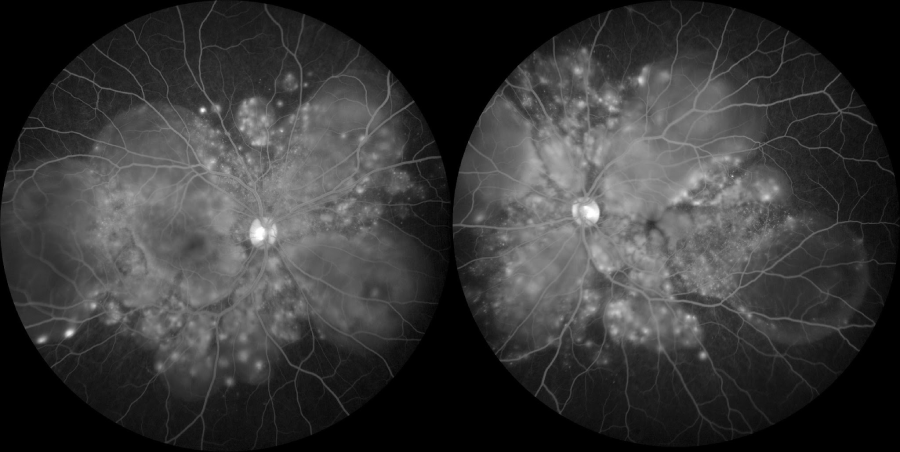
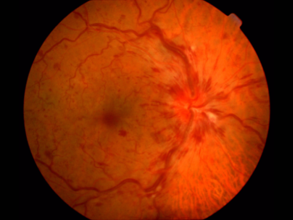
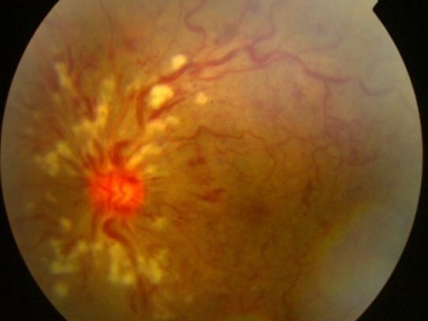
Retinitis pigmentosa with consecutive optic atrophy
Patients with night blindness, Progressive visual deterioration, Positive family history, Progeny of a first degree consanguinous Marriage
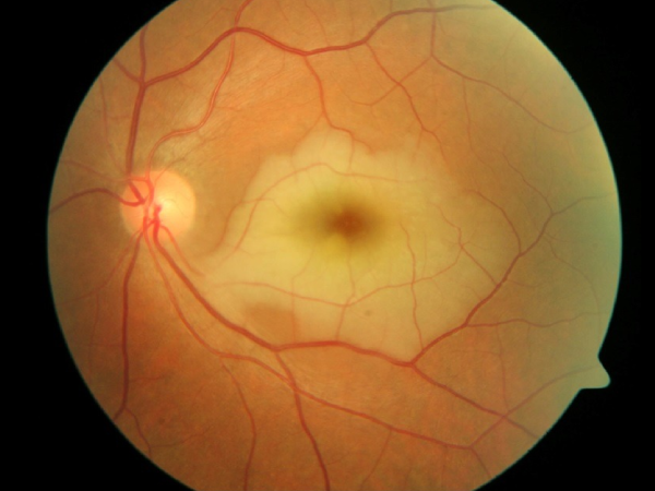
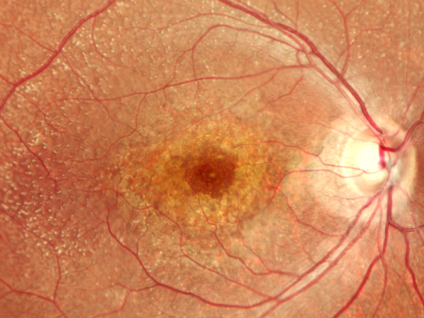
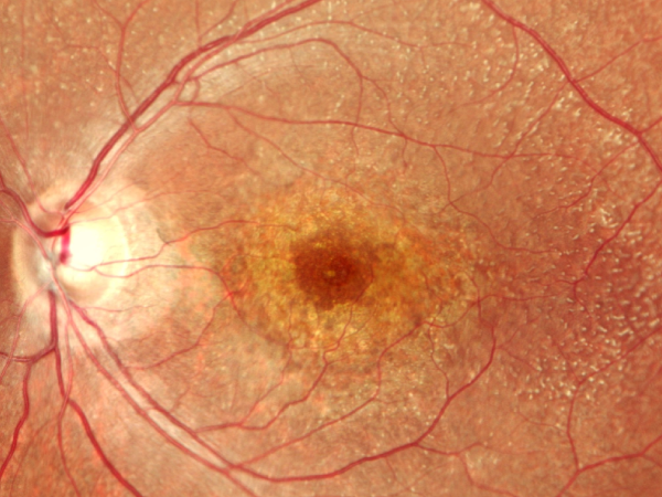
Heredomacular dystrophy presents with subacute onset of progressive central visual loss
Symptoms of Retinal Disorders
Sudden or gradual vision loss
May affect central or peripheral (side) vision and can progress quickly in serious conditions like retinal detachment.
Floaters
Appear as black spots, cobwebs, or threads moving across your field of vision, often linked to changes in the vitreous gel or retinal tears.
Flashes of light
Brief flashes, especially in dim environments, may signal traction on the retina and risk of detachment.
Blurry or distorted central vision
Common in macular conditions like macular degeneration or holes.
Shadow or curtain across vision
Often described as a dark veil moving from one side; an urgent sign of retinal detachment.
Night vision problems
Difficulty seeing in dim light can indicate early retinal dysfunction.
Straight lines appear wavy (metamorphopsia)
Suggests macular involvement, often seen in macular degeneration or epiretinal membranes.
Causes & Risk Factors
Diabetes Mellitus
Chronically high blood sugar can damage tiny retinal vessels, leading to diabetic retinopathy.
High Blood Pressure
Affects retinal circulation, increasing the risk of retinal vein occlusion or bleeding.
Aging
Natural wear of retinal tissue increases the chance of macular degeneration or macular holes.
High Myopia (Nearsightedness)
Elongated eyeballs in severe myopia stretch and thin the retina, raising the risk of tears and detachment.
Eye Injuries or Surgeries
Trauma or previous procedures like cataract surgery may alter the vitreous or retinal structure.
Genetic Factors
Family history plays a role in age-related macular degeneration (AMD) or inherited retinal dystrophies.
Poor Lifestyle Choices
Smoking and nutrient-deficient diets accelerate retinal degeneration, especially in AMD.
Vitreo-Retinal Diagnostic Technology
To provide accurate, early detection and targeted care, Chakrabarthi Eye Care Centre employs advanced retinal imaging and diagnostic tools:
Optical Coherence Tomography
High-resolution imaging of the retina and macula for structural analysis.
Fundus Fluorescein Angiography
Visualizes blood flow in the retina to detect leakage, blockage, or neovascularization.
B-Scan Ultrasonography
Evaluates the posterior segment in cases with media opacity.
Wide-Field Retinal Imaging
Captures ultra-wide views of the peripheral retina, essential for diabetic or degenerative cases.
OCT Angiography
A non-invasive method to assess blood flow in retinal capillaries, ideal for AMD or diabetic eyes.
Vitreo-Retinal Treatment Options
Intravitreal Injections
Anti-VEGF drugs (e.g., Avastin, Eylea, Lucentis) are injected directly into the eye to block abnormal vessel growth and reduce leakage in diseases like AMD and diabetic retinopathy.
Laser Photocoagulation
A precise laser is used to seal leaking blood vessels or prevent further retinal damage in cases of diabetic retinopathy and vein occlusion.
Pars Plana Vitrectomy
A microsurgical procedure to remove the vitreous gel and repair retinal tears, detachments, macular holes, or severe hemorrhages. Advanced techniques like 23G/25G/27G sutureless vitrectomy offer faster recovery.
Pneumatic Retinopexy
Used in selected retinal detachment cases, a gas bubble is injected into the eye to push the retina back in place, followed by laser or cryotherapy to seal the tear.
Scleral Buckling
An external band is placed around the eye to support the retina from outside and relieve traction caused by retinal tears. Commonly used in younger patients.
Our Retina Specialists
Our experienced vitreo-retinal surgeons bring extensive expertise in managing diabetic eye disease, macular conditions, and complex retinal surgeries. With a patient-focused approach and access to the most sophisticated diagnostic and therapeutic tools, they ensure safe, effective outcomes tailored to individual visual needs. From timely injections to advanced vitrectomy techniques, our retina team is committed to preserving sight and enhancing quality of life.