
Vitreo-Retinal Surgery
Retina surgery is performed to repair and restore vision affected by retinal disorders such as retinal detachment, macular hole, or diabetic retinopathy. Using advanced microsurgical techniques, the surgeon carefully addresses the underlying condition to preserve and improve eyesight.
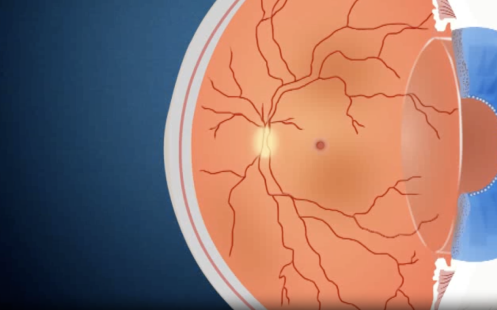
Vitreoretinal surgeries are a group of delicate procedures performed inside the eye to address problems affecting the retina, macula, and vitreous gel. These intricate operations are used to treat a wide variety of serious eye conditions and help restore, preserve, or improve vision.
Benefits & Advantages
- Restores and preserves vision in retinal conditions
- Prevents further vision loss or blindness
- Uses advanced, minimally invasive surgical techniques
- Improves overall quality of life and daily functioning
Common procedures and conditions treated
The type of vitreoretinal surgery performed depends on the specific condition and location of the problem.
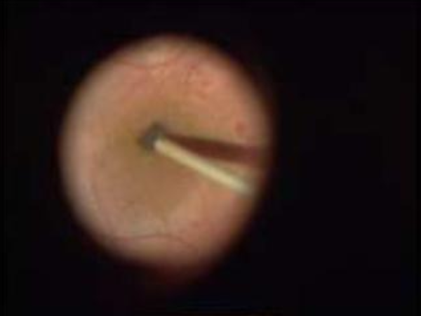
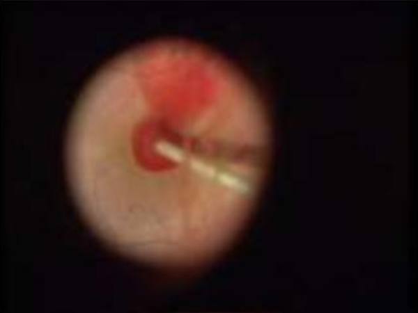
Vitrectomy For Diabetic Retinopathy Surgery
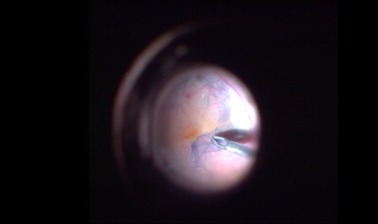
Vitrectomy
This procedure involves removing the vitreous humor, the gel-like substance that fills the center of the eye. Reasons for a vitrectomy include:
Diabetic retinopathy
Clearing blood and scar tissue caused by damaged blood vessels.
Vitreous hemorrhage
Removing blood that has seeped into the vitreous gel.
Retinal detachment
Gaining access to the retina to repair tears or detachment.
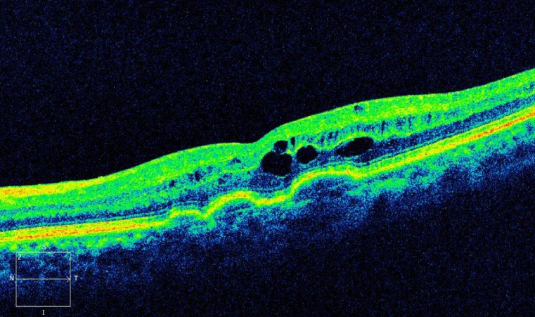
Macular holes and puckers
Repairing holes or peeling away scar tissue (epiretinal membranes) from the macula.
Trauma
Removing foreign objects from the eye.
Infection
Treating endophthalmitis, a severe eye infection.
Retinal Detachment Repair
If the retina has detached, one or more surgical techniques may be used in combination
Scleral buckle
A permanent band, usually made of silicone, is sewn onto the white of the eye (sclera). This gently pushes the sclera inward, forcing the retina to reattach.
Pneumatic retinopexy
An expanding gas bubble is injected into the eye. The patient is positioned so that the bubble presses against the detached retina, holding it in place while it heals.
Retinal cryopexy
An intense freezing probe is used to scar the tissue around a retinal tear, sealing it and helping to prevent further detachment.
Laser photocoagulation
A laser is used to create scars that weld the retina to the underlying tissue.
Retinal Detachment with PVR
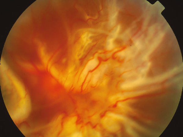
Retinal Detachment with Large Posterior Tear and Macular Hole
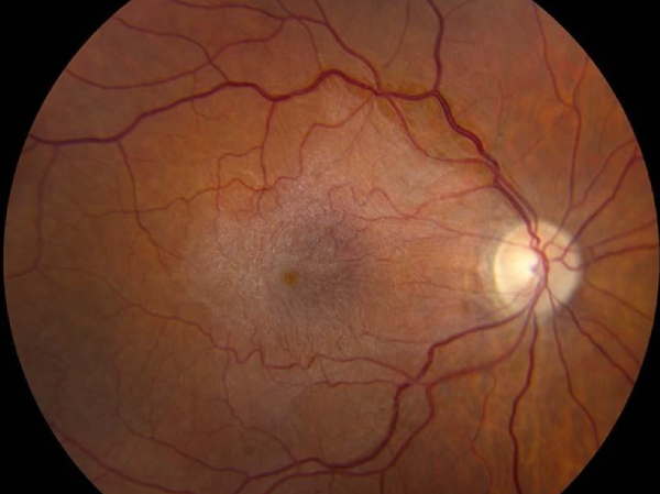
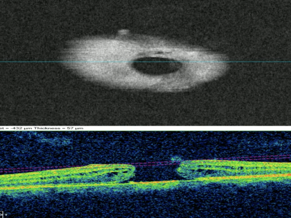
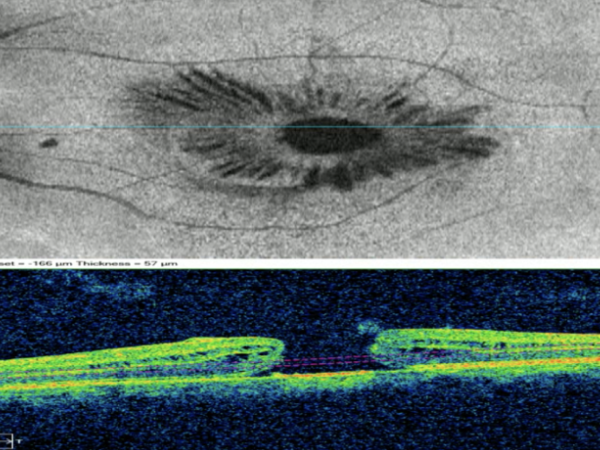
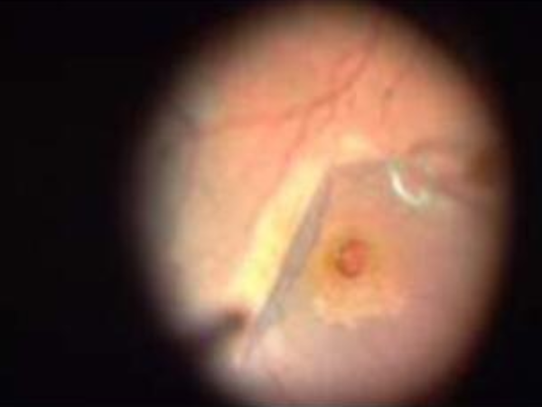
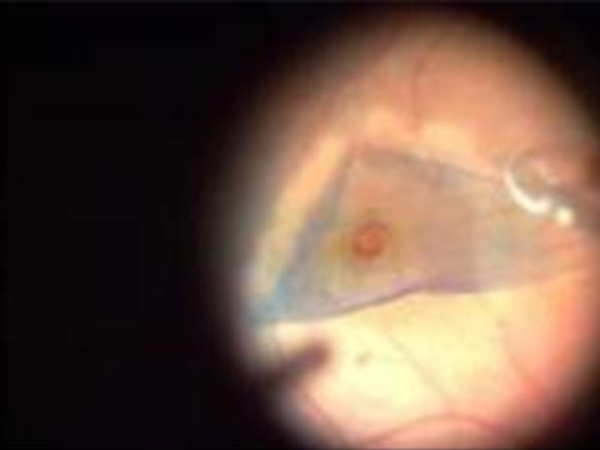
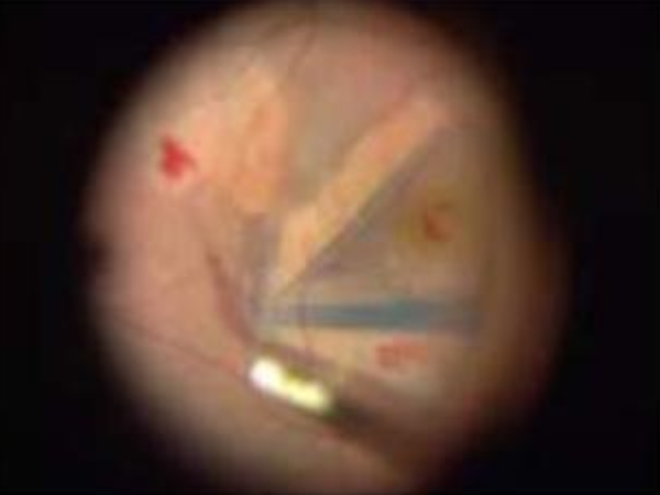
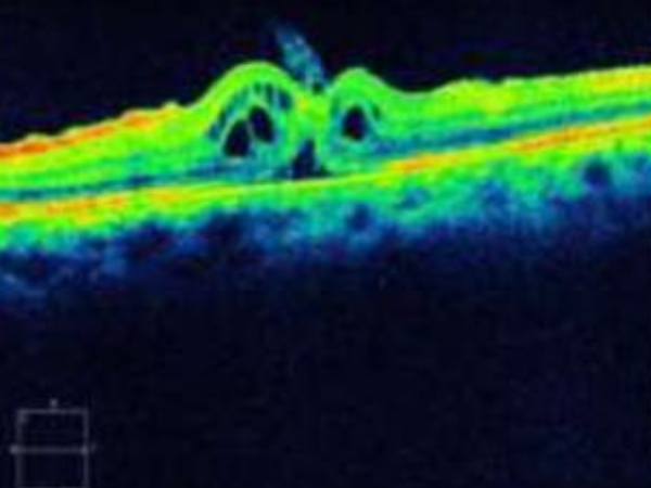
Macular Hole Repair
This procedure is used to close a hole in the macula. A vitrectomy is first performed, after which the surgeon peels a thin membrane from the retinal surface called the internal limiting membrane and uses that membrane along with the patient’s own autologous blood to form a plug to close the hole.
Intravitreal air or gas tomponade s not necessary for the MACULAR PLUG technique. also these patients do not need prone positioning and there is no restriction to air travel after this procedure
The surgical process
Anesthesia
Vitreoretinal surgery can be performed under local or general anesthesia, depending on the patient's condition and the complexity of the procedure.
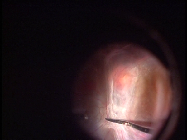
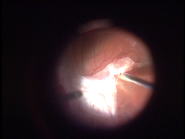
Instruments
A surgeon uses a microscopic cutting device (vitrector), fiber-optic light, and other miniature instruments to operate inside the eye through three tiny incisions.
Recovery
Patients may use antibiotic and anti-inflammatory eye drops for several weeks. Vision will be blurry at first, with final results taking a few weeks to several months to be realized.
Factors affecting success and recovery
The success rate of vitreoretinal surgery is generally high, with vision significantly improved or restored for many patients. However, outcomes can depend on:
The underlying condition
A detached retina, for example, is best treated quickly before the detachment spreads to the macula.
Patient factors
The patient's overall health and age can influence recovery.
The specific procedure
More complex cases, such as proliferative vitreoretinopathy (PVR), can have a longer recovery and lower visual prognosis.
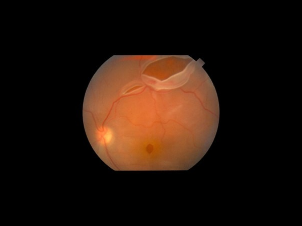
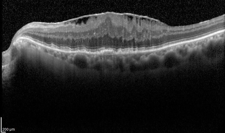
Vitrectomy for Macular Hole
Postoperative After Vitrectomy and
ERM Peeling
ERM /Epiretinal membranes will cause defective vision both for distance & near and metamorphopsia.This condition requires surgery in symptomatic patients
Vitrectomy for Epiretinal
Membrane
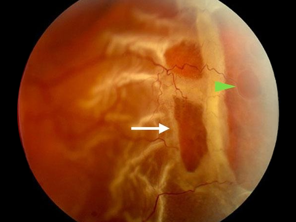
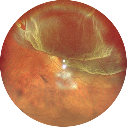
Symptomatic posterior vitreous detachment
with large horseshoe tear
Patients with floaters and flashes need a detailed dilated fundus examination

Scleral Buckling
Scleral buckling for rhegmatogenous retinal detachment Combined with pars plana vitrectomy and gas/oil Tamponade is the treatment of choice