Fluorescein Fundus Angiography (FFA)
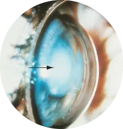
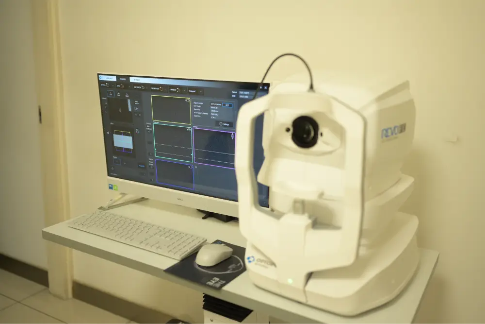
Fluorescein Fundus Angiography is a specialized diagnostic procedure that uses a fluorescent dye and a camera to capture detailed images of the blood vessels in the retina and choroid at the back of the eye. This test helps ophthalmologists diagnose and monitor retinal vascular diseases and other conditions affecting the eyes circulation.
Uses
- Diagnosing and monitoring diabetic retinopathy
- Evaluating age-related macular degeneration (AMD)
- Detecting and assessing retinal vein or artery occlusions
- Identifying choroidal neovascularization and other abnormal blood vessel growth
- Planning and monitoring treatment for retinal diseases such as macular edema
Benefits & Advantages
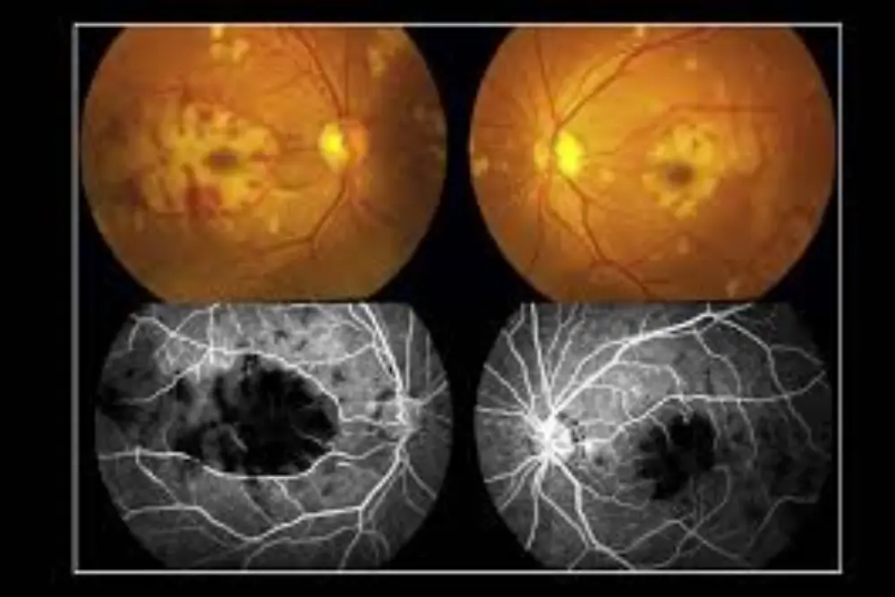
- Provides high-resolution images of retinal and choroidal blood flow
- Helps detect blood vessel abnormalities not visible during routine exams
- Essential for diagnosing and managing serious retinal diseases
- Guides targeted treatment such as laser therapy or injections
- Enables early intervention to prevent vision loss
Why Fluorescein Fundus Angiography is Important
FFA offers a unique window into the health of the retinal circulation, enabling early diagnosis and precise management of sight-threatening diseases. By visualizing blood flow abnormalities, ophthalmologists can tailor treatments that preserve vision and improve outcomes.