Anterior & Posterior Segment OCT

Anterior Segment OCT

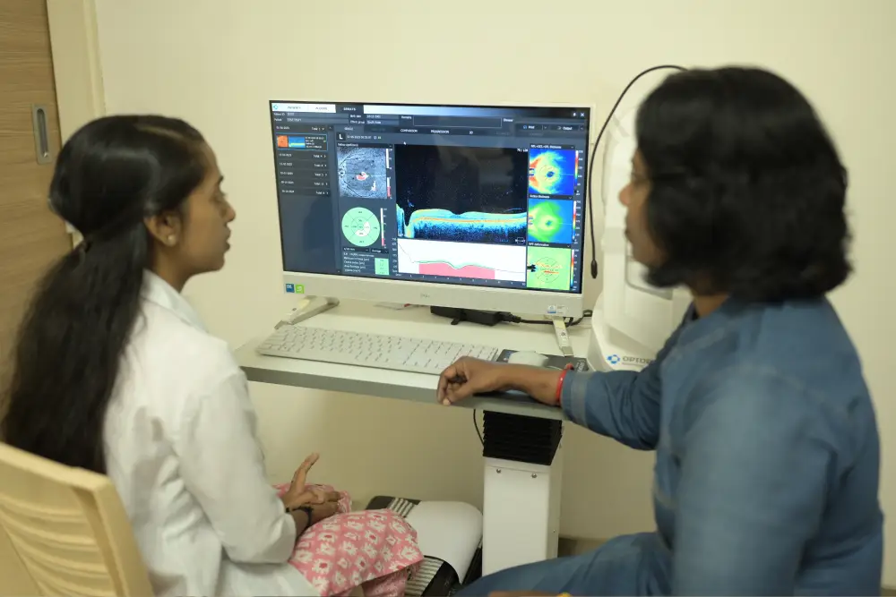
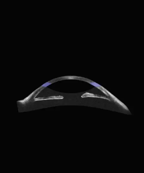
Anterior Segment OCT focuses on imaging the cornea, iris, anterior chamber, and lens. It is particularly useful in evaluating conditions affecting the front structures of the eye.

Uses

  • Measuring corneal thickness (pachymetry)
  • Diagnosing and monitoring keratoconus
  • Assessing angle-closure glaucoma
  • Evaluating corneal grafts and refractive surgery outcomes
  • Measuring anterior chamber depth for lens implant planning
  • Imaging ocular surface tumors or lesions

Benefits & Advantages

  • Non-contact, quick, and painless
  • Detects subtle changes in corneal structure
  • Provides precise measurements for surgical planning
  • Essential for glaucoma diagnosis and management
  • Guides refractive and cataract surgery decisions

Posterior Segment OCT

Posterior Segment OCT images the retina, macula, and optic nerve head, providing detailed cross-sectional views of these delicate structures.

Uses

  • Diagnosing and monitoring macular degeneration
  • Detecting macular holes, cysts, or edema
  • Managing diabetic retinopathy
  • Monitoring glaucoma progression (optic nerve analysis)
  • Assessing retinal detachment or epiretinal membranes
  • Evaluating vitreomacular interface disorders

Benefits & Advantages

  • High-resolution imaging for early detection of retinal diseases
  • Monitors treatment response in conditions like macular edema
  • Guides retinal surgery planning
  • Detects changes invisible to standard examination
  • Non-invasive and safe for repeated use
Book Appointment Book Appt. Call Now Doctors Whatsapp